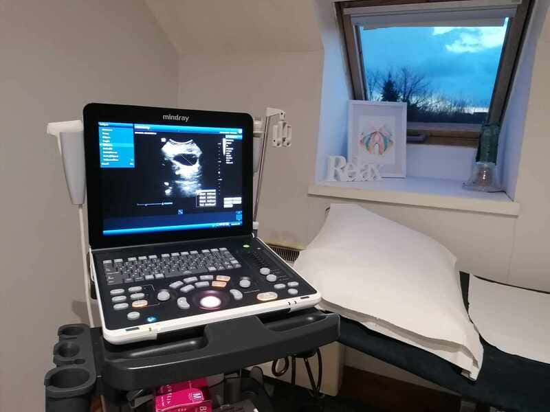
A pelvic ultrasound is an imaging exam that creates a picture of your pelvic organs in real-time.
It is used in addition to a pelvic floor/abdominal examination, exercises and tips, essentially as a visual biofeedback tool. It is not used as a medical diagnosis (neither as foetal scan).
As described below, the ultrasound can be used to assess your pelvic organs, pelvic floor and abdominal muscles.
The examination is non-invasive and painfree, using a monitor, a probe and some gel and following strictly all the hygiene recommendations.
Assessing your bladder is essential if you present urinary symptoms as urgency, frequency, UTI’s, retention or loss of bladder control.
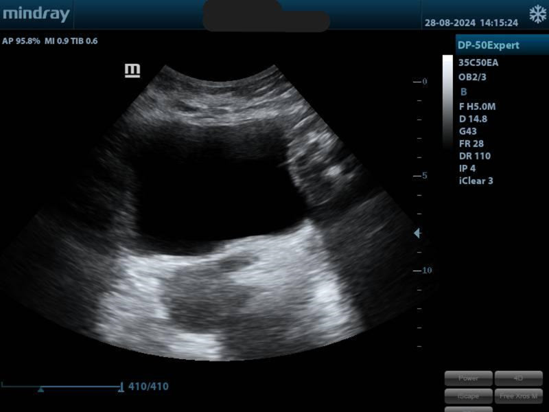
When placing the probe on the lower tummy, we can get an image of the bladder.
We will check if the shape and volume are normal. We will measure your bladder before and after urination, checking if any residue subsists. We can also assess its support when coughing, pushing and contracting the pelvic floor muscles.
Depending on how your bladder looks, we might change your bladder habits: drinking more water, urinating more or less often, emptying your bladder completely, knacking, …

Another way to check your pelvis area is with a trans-perineal view (the probe will be placed on your perineum). It will give us a picture of your internal organs:
You will be able to see where your organs are, what happens when you contract your pelvic floor, when you hold or relax it, when coughing, pushing, laughing, moving… and if there is any sign of prolapse.
A visual feedback can help to understand better what you’re doing.
This examination can be performed in different positions as it can be different lying or standing.
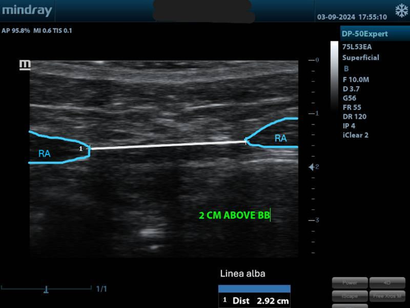
The tummy check can be very interesting especially in post-partum, after abdominal surgery or if you suspect having a diastasis recti (separation between your abs).
We will:
